
Figure 1.
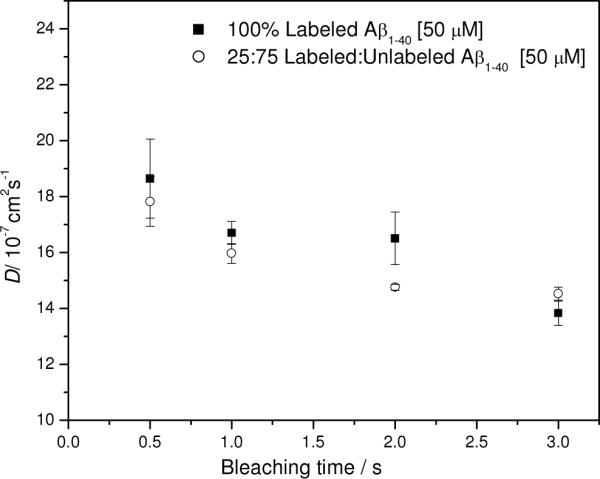
Diffusion as a function of bleaching time for labeled Aβ(1–40) and a mixture of labeled and unlabeled Aβ(1–40). Filled squares: 100% 5-carboxyfluorescein Aβ(1–40), 50 μM peptide. Open circles: mixture of 25% 5-carboxyfluorescein-labeled Aβ(1–40) with 75% Aβ(1–40) expressed as percentage of 50 μM peptide. Conditions: 50 mM PBS, 150 mM NaCl, pH 7.4, 0.5 Watts laser power. Error bars represent standard deviations from triplicate runs.