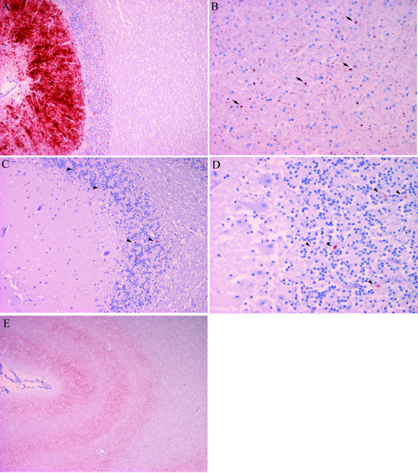
Figure 2.
Immunohistochemistry for PrPSc using a cocktail of mAb F89/160.1.5 and 2G11. A) Cerebellum of case M72 (100×) showing intense immunostaining in the molecular layer of cerebellar cortex. B) Frontal lobe of case M27 with F89/160.1.5/2G11, (400×) showing punctate globular immunostaining (arrow) in white matter. C) PrPSc deposits (arrowheads) in the granular layer of the cerebellum of case M27. Scant fine granular deposits of PrPSc visible in the granular layer (200×). D) Cerebellar cortex of case M15 with scant fine granular (arrowheads) deposits of PrPSc visible in the granular layer (400×). E) Cerebral cortex of case M15 (40×) showing fine granular immunolabelling following a laminar pattern.

