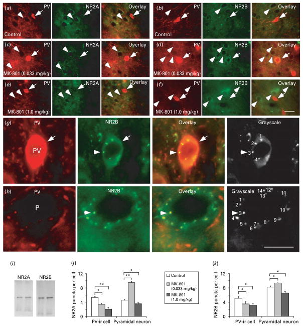
Fig. 5.
MK-801-induced distinct changes of NR2A and NR2B subunits in parvalbumin (PV)-containing interneurons vs. pyramidal neurons. (a–f) Representative photographs of double immunofluorescent labelling of PV (red) and NMDAR subunits (green) in controls (a, b) and MK-801 treatment at low dose (c, d) and high dose (e, f). The PV-immunoreactive (ir) interneurons (arrows) were double-labelled by PV and NR2A or NR2B, whereas the putative pyramidal neurons (arrowheads) were surrounded by both PV-ir axon terminals (red) and labelled NR2 positive puncta (green). Scale bar in (f)=20 μm for all images in panels (a)–(f). (g, h) Images at high magnification showing the methods used for quantification of NR2A and NR2B puncta in both PV-ir cells (g) and pyramidal neurons (h). Scale bar in (h)=20 μm for all images in panels (g) and (h). (i) Western blot analyses showing the specificities of anti-NR2A and anti-NR2B used for immunostaining. Both antibodies exhibited single molecular weight band. (j, k) Summary histograms showing changes of NR2A and NR2B subunit protein expression (puncta numbers) in the PV-ir interneurons and pyramidal neurons in MK-801-treated rats. Compared to control, 0.033 mg/kg MK-801 treatment significantly decreased the expression of both NR2A and NR2B subunits in PV-ir cells (p<0.05). In contrast, MK-801 significantly increased the puncta numbers of both NR2A and NR2B subunits in pyramidal neurons at low dose (0.033 mg/kg) but dramatically decreased the puncta numbers at high dose (1 mg/kg), suggesting distinct cell-type-specific effects in PFC circuitry (* p<0.05, ** p<0.01).

