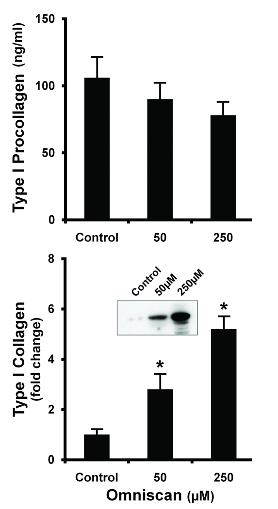
Figure 1. Production of type I procollagen and deposition of type I collagen in the cell layer in Omniscan-treated cells.
Cells were incubated under control conditions or in the presence of Omniscan for three days. At the end of the incubation period, cell culture fluid was collected and assayed for type I procollagen by ELISA (Upper panel). Lysates were prepared from the cell layer (Lower panel) and assayed for type I collagen by western blotting. Values shown are means and standard errors based on n=5 separate experiments. The inset showing intact type I collagen is from one of the replicate experiments with β-tubulin (no change) used as control. Statistical significance of the data was assessed by ANOVA, followed by paired-group comparisons. *indicates statistically significant increase compared to control at p<0.05 level.

