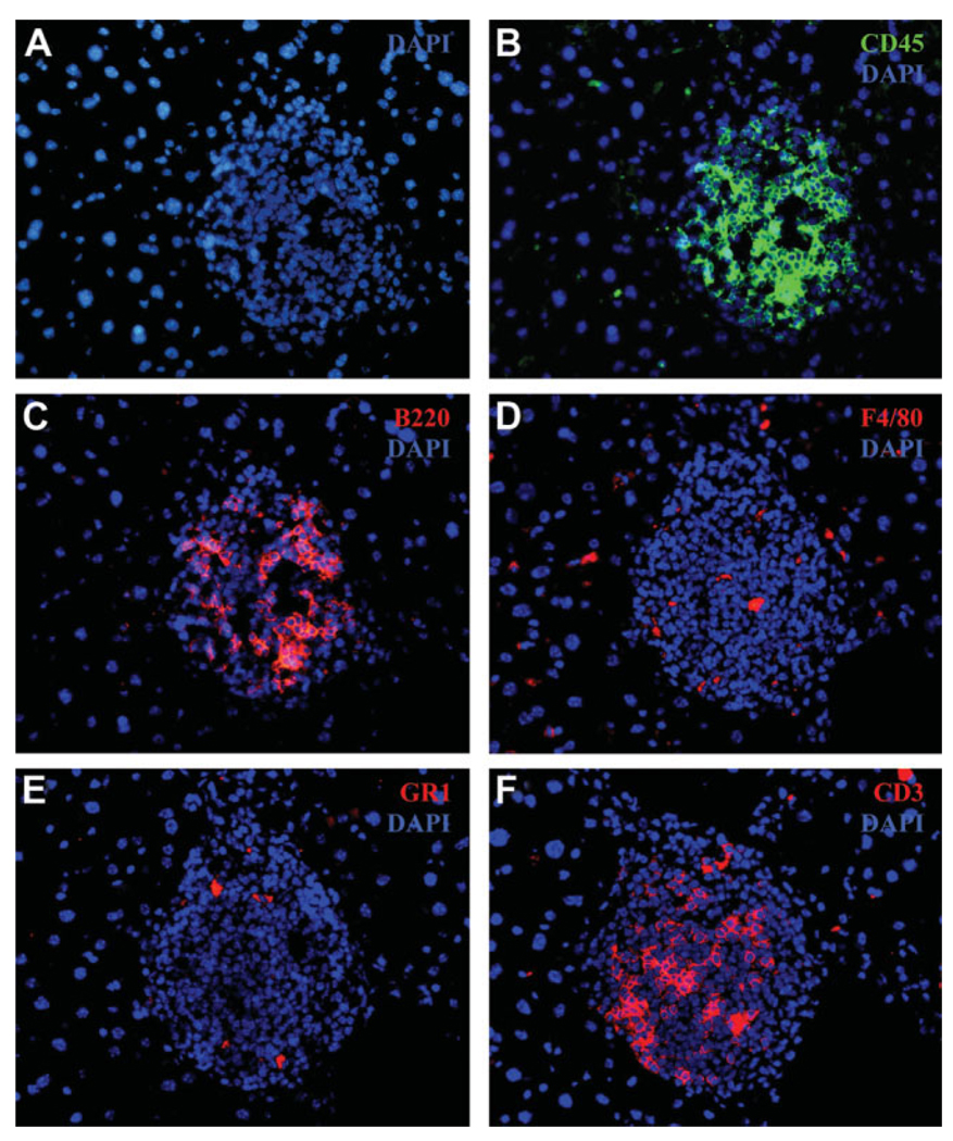
Fig. 3.
Frozen sections from old liver tissue (24 months) were paraformaldehyde-fixed and stained with (A) nuclear DAPI stain (blue) and the following immune cell markers: (B) CD45 for leukocytes, (C) B220 for B cells, (D) F4/80 for macrophages, (E) GR1 for granulocytes (neutrophils), and (F) CD3 for T cells (total). Hepatocytes can be visualized with DAPI as larger, round, isolated nuclei, whereas the immune cell cluster can be seen as a region of denser concentration of DAPI staining smaller nuclei (magnification ×40).

