Fig. 3.
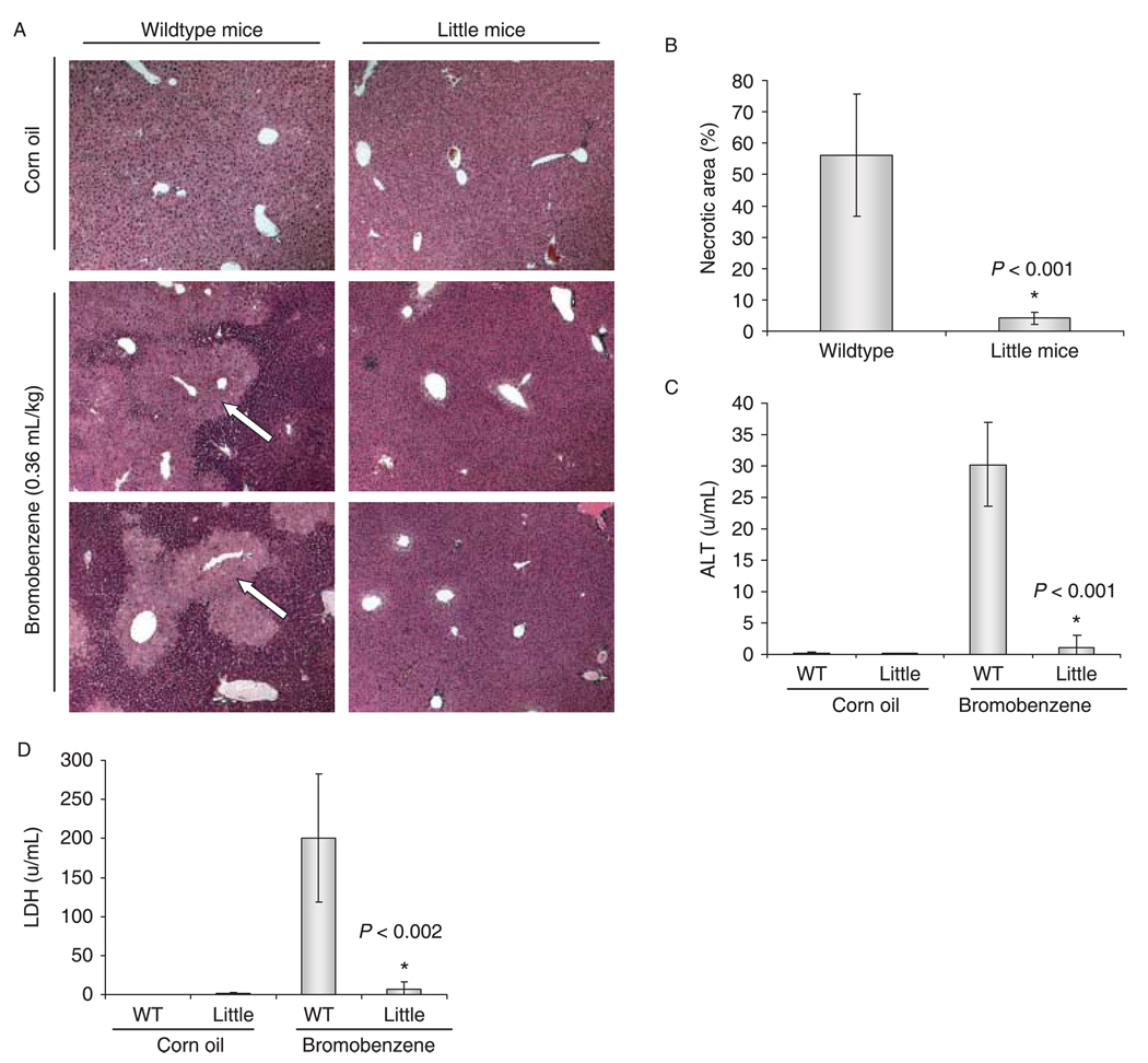
Little mice are resistant to bromobenzene-induced liver toxicity. Mice were treated with a 0.36 mL kg−1 dose of bromobenzene by intraperitoneal injection. (A) After 24 h, liver sections were examined by histological staining (hematoxylin and eosin staining). Liver samples for all treated animals were analyzed and representative histology is shown. The livers from wild-type animals (n = 5) showed extensive necrosis (indicated by arrows), but only minimal necrosis was observed in Little mice (n = 5). (B) The necrotic area (as a percentage of total area) was determined for each experimental group as described in the methods section. Little mice showed a significantly reduced necrotic area as compared to wild-type mice (P < 0.001). Serum levels of ALT (C) and LDH (D) were measured 24 h after treatment. Little mice showed significantly reduced levels of both ALT (P < 0.001) and LDH (P < 0.002). In (B), (C), and (D) bars represents the mean ± (2 × SE) for each group. All P-values refer to a Student’s t-test.

