
Figure 4.
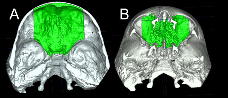
Anatomic structures used for superimpositions of 3D models of growing subjects. The anterior cranial fossa region of the cranial base 3D surface models after treatment was used for registration A shows the superior view and B the inferior view