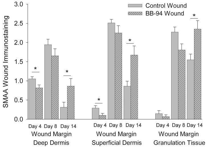
Figure 3.
Semiquantitative analysis of the SMAA immunostaining in superficial and deep dermis of wound margins and in granulation tissue of control and BB-94 treated wounds. SMAA immunostaining in superficial and deep dermis of wound margins and in granulation tissue was assessed semiquantitatively on a four-graded scale: 0 = no positive staining, 1 = weak, 2 = moderate and 3 = abundant. BB-94 treatment of wounds resulted in reduced SMAA immunostaining on day 4 and 8 and increased immunostaining on day 14, compared to control wounds. Control, open bars; BB-94, filled bars. *p<0.05. Mean ± SEM. n=12 on days 4 and 14, n=9 on day 8.

