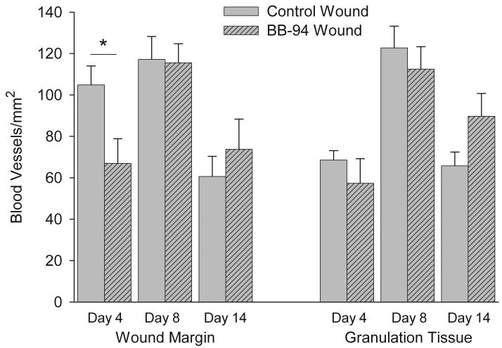
Figure 4.
Quantitative analysis of number of blood vessels in superficial and deep dermis of wound margins and in granulation tissue of wounds. Blood vessels were identified by SMAA immunostained perivascular cells surrounding a lumen and counted as described in Materials and Methods. BB-94 treated wounds were delayed in initial blood vessel formation and in the later reduction in blood vessel numbers. Control, open bars; BB-94, filled bars. *p<0.05. Mean ± SEM. n=12 on days 4 and 14, n=9 on day 8.

