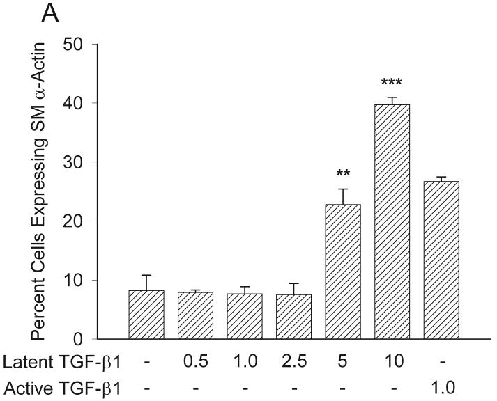
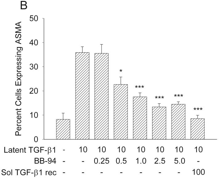
Figure 8.
Expression of SMAA in response to TGF-β1 with or without BB-94. (A) Latent TGF-β1significantly induced myofibroblast formation at concentrations of 5 and 10 ng/ml to levels similar to those of active TGF-β1, as assessed by increased expression of SMAA **p<0.01; ***p<0.005. Mean ± SEM. (B) Addition of increasing concentrations of BB-94 (0.5-5.0 μM) significantly inhibited the ability of latent TGF-β1 to promote SMAA expression. The soluble TGF-β receptor antagonist significantly inhibited latent TGF-β1-promoted SMAA expression. The percentage of cells expressing SMAA was determined by counting at least 100 cells per treatment group from three treatment groups. Each experiment was repeated at least three times. *p<0.05; ***p≤0.001. Mean ± SEM.


