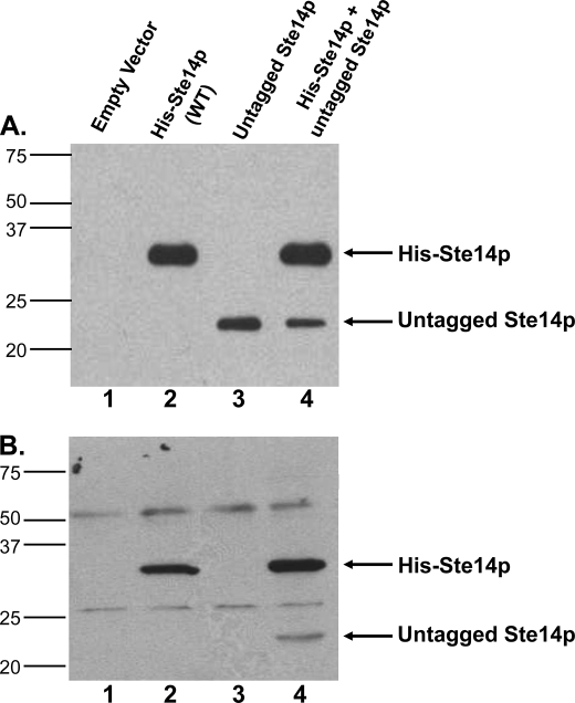
FIGURE 2.
Expression and co-immunoprecipitation of untagged Ste14p with His-Ste14p. A, 2 μg of crude membrane protein expressing tagged or untagged Ste14p variants were subjected to 10% SDS-PAGE and immunoblot analyses. First lane, empty vector; second lane, His-Ste14p; third lane, untagged-Ste14p; fourth lane, His-Ste14p + untagged-Ste14p. Proteins were detected with a Ste14p polyclonal antibody (1:500) and HRP-conjugated goat anti-rabbit secondary antibody (1:10,000), and the bands were visualized using ECL. B, 100 μg of protein from crude membrane preparations were incubated in 1× RIPA buffer plus ∼3 μg of anti-Myc monoclonal antibody overnight at 4 °C with gentle rotation. 40 μl of 50% (vol:vol) protein A-Sepharose beads in 1× RIPA buffer were added to each tube and incubated another 2.5 h at 4 °C with gentle rotation. The bead-immuno-protein complexes were processed as described under “Experimental Procedures,” and the entire sample was separated by 10% SDS-PAGE. Immunodetection was with a Ste14p polyclonal antibody (1:500) and a goat anti-rabbit secondary antibody (1:10,000) conjugated to HRP. Protein bands were visualized by ECL. WT, wild type.

