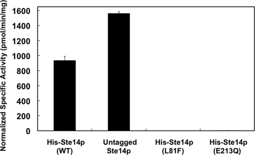
FIGURE 4.
In vitro methyltransferase activities of Ste14p variants in crude membranes. 5 μg of crude membrane protein were incubated with 200 μm AFC and 20 μm [14C]SAM for 30 min at 30 °C in a total volume of 60 μl. The reactions were terminated by the addition of 50 μl of 1 m NaOH/1% SDS (v/v), and the activity was quantified by the vapor diffusion assay as described under “Experimental Procedures.” Each reaction was performed three times in duplicate, and error bars represent ±S.D.). Activities were normalized to the expression level of each construct using immunoblots containing varying amounts of each protein. The protein bands were visualized with ECL as described in Fig. 3 and quantified using a Nucleovision imaging system equipped with a high sensitivity cooled CCD camera. WT, wild type.

