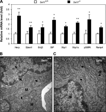
FIGURE 2.
Sel1l−/− embryos develop systemic endoplasmic reticulum stress. A, quantitative analysis of ER stress-responsive genes in Sel1l−/− embryos. Total RNAs were isolated from Sel1l+/+ and Sel1l−/− embryos at E12.5. The mRNA expression of several ER stress-inducible genes in Sel1l+/+ and Sel1l−/− embryos was analyzed by quantitative RT-PCR. Data are expressed as expression-fold differences between Sel1l+/+ (set to 1) and Sel1l−/− embryos. n = 3 embryos per genotype, *, p < 0.05; **, p < 0.01 mutant versus wild-type control. B and C, electron micrographs of liver sections from Sel1l+/+ and Sel1l−/− embryos at E12.5. Arrows indicate endoplasmic reticulum; N, nucleus; M, mitochondria. Note that Sel1l−/− hepatocytes exhibit a significantly dilated ER (arrows in C) relative to those of Sel1l+/+ hepatocytes (arrows in B).

