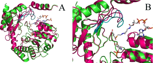
FIGURE 1.
The position of the active site loop in the R. capsulatus ALAS crystal structure. In A, the ribbon representations of the three-dimensional structures of one monomer of holoenzymic (magenta) and succinyl-CoA-bound (green) ALAS from R. capsulatus are superimposed. Note the distinct positioning of the active site loop in the two structures (depicted in magenta in the ALAS holoenzyme structure and in teal in the succinyl-CoA-bound structure). In B, the active site loop in the closed conformation of ALAS (i.e. succinyl-CoA-bound structure) is perched above the catalytic cleft of the enzyme. Succinyl-CoA and the cofactor PLP are shown as sticks. The image was constructed using Pymol and Protein Data Bank entries 2BWN and 2BWO.

