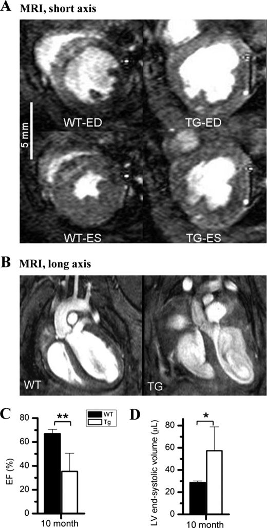
FIGURE 3.
MRI. Short axis MRI images show a systolic dysfunction represented by a significant increase in end systolic (ES) volume and significantly decreased LV ejection fraction due to decreased % fractional shortening in the TEAD-1 transgenic heart (A). Long axis MRI images reveal a change in the morphology of 10-month-old TEAD-1 Tg line 12 hearts (B). Significantly decreased LV ejection fraction (EF) (C) and significantly increased end systolic (ES) volume (D) are shown. (n = 5). *, p < 0.05; **, p < 0.01. ED, end diastolic labeling.

