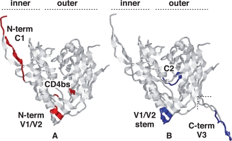
FIGURE 8.
Location of the interaction sites for PDI and CNX in the mature gp120 structure. The gp120 structure shown was rendered from the recently released structure (Protein Data Bank code 3JWO (50)) that includes an extended N terminus when compared with earlier structures. The V3 loop in B was rendered from the single structure reporting it (Protein Data Bank code 2B4C) and then grafted where shown. The rendering is in the canonical view of gp120 (45) with the inner and outer domains of the molecule indicated. The definitive epitopes for the Abs that blocked chaperone binding are indicated, where available. PDI-reactive sites are largely accessible on the mature molecule (A), and those reacting with CNX are buried or face inward (B), consistent with access only prior to folding. The sites are mutually exclusive except for the V1/V2 region where precise residues cannot be indicated as they are not present in any solved structure. The diagram is illustrative, and the actual structure of the molecule during interaction with either chaperone remaining unknown.

