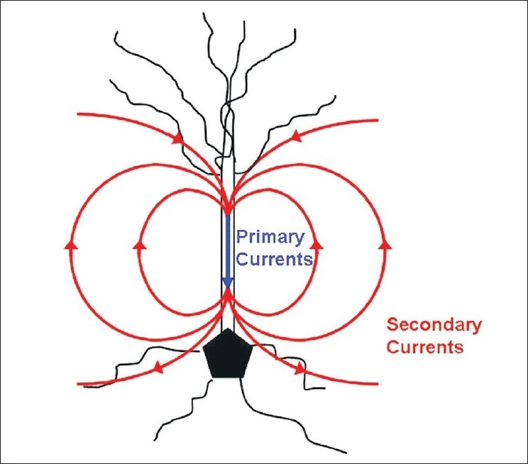
Figure 1.

A neuronal pyramidal cell is seen in this image with primary (intracellular) currents and secondary (volume/extracellular) currents. Primary currents are depicted in blue and secondary currents in red. MEG signals are a measure of the intracellular current produced by the apical dendrites and therefore more apt to accurately represent the actual source generator. EEG signals recorded at the scalp electrodes are a measure of extracellular currents
