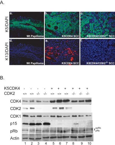
Figure 4.
Keratin 13 expression and biochemical analysis of mouse skin tumors. (A) Expression of normal differentiation marker keratin 5 (K5) (green) was detected by immunofluorescence in all paraffin cross sections of wild type papillomas (a), K5CDK4 SCC (b), and K5CDK4/CDK2−/− SCC (c). Note that while the normal pattern of K5 expression (basal cell layer), as seen in wt papillomas, is lost in SCCs. SCC from K5CDK4 mice (e) stain positive for keratin 13 (K13) (red), marker associated with malignant progression. Magnifications at 20X; Dapi (blue) used as nuclear counter stain.
(B) Protein lysates from 30 week papillomas obtained from wt (lines 1, 2), CDK2−/− (lines 3, 4), K5CDK4 (lines 5, 6, 7) and K5CDK4/CDK2−/− (lines 8, 9, 10) mice were separated by SDS-PAGE, transferred to nitrocellulose membrane and blotted for CDK4, CDK2, CDK1, p15Ink4b and pRb. Actin was used as loading control. Hyperphosphorylation (ppRb) and hypophosphorylation (pRb) of retinoblastoma protein was denoted on the right.

