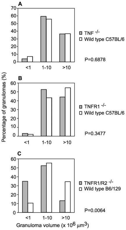
Fig. 1.
Size distribution of early granulomas in TNFR1/R2−/−, TNFR1−/− and TNF−/− mice. Livers from Schistosoma mansoni-infected TNF−/− (A), TNFR1−/− (B) and TNFR1−/−/R2−/− (C) mice at 6 weeks p.i. were sectioned and stained with H&E. Sections were systematically scanned and all granulomas with a visible central egg were measured with an ocular micrometer to determine granuloma volume. Granulomas were then assigned, depending on volume, to one of three categories: <1 × 10⁶, 1–10 × 106 and >10 × 106 μm3. Differences in the distribution of granuloma volumes in each genotype when compared to the appropriate wild type mice were assessed using χ2 tests and the P values returned by these tests are indicated.

