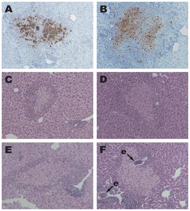
Fig. 2.
Hepatocyte apoptosis in Schistosoma mansoni-infected TNFR1/R2−/−, TNF−/− and TNF/LT−/− mice. Liver sections from S. mansoni-infected wild type (A, C), TNFR1−/−/R2−/− (B, D), TNF−/− (E) and TNF−/−/LT−/− (F) mice were stained by the TUNEL method (A, B) or with H&E (C–F). Apoptotic nuclei in A and B are stained dark red. Areas of apoptotic hepatocytes in C–F appear paler and slightly more eosinophilic than nearby healthy tissue, and are surrounded by smaller, darker-staining inflammatory cells. e, schistosome egg. 100 × magnification.

