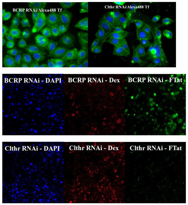
Fig. 3.
Effective clathrin heavy chain RNAi eliminated both the punctate and the diffused intracellular fluorescence of Flu-R.I.-CKTat9 in live HeLa cells. HeLa cells were double transfected with the negative control BCRP siRNA or the clc-2 siRNA, incubated with 10 μg/ml of DAPI and 0.35 μM Alexa 488-Tf for the fluorescence microscopy (top two images, respectively, captured with a 63× objective) or 10 μg/ml of DAPI, 75 μM Rhodamine-Dextran (Dex), and 10 μM of Rho-R.I.-CKTat9 (FTat) for the confocal microscopy (middle and bottom six images, respectively), washed, and examined microscopically. BCRP RNAi/Alexa488 Tf shows abundant punctate Tf fluorescence, which is absent in Clthr RNAi/Alexa488 Tf. BCRP RNAi-DAPI, BCRP RNAi-Dex, and BCRP RNAi-FTat are confocal blue, red, and green channel images, respectively, of the same middle section of a field captured using a 40× objective, so are Clthr RNAi-DAPI, Clthr RNAi-Dex, and Clthr RNAi-FTat. Comparison between the BCRP and the Clathrin RNAi images shows that Clathrin heavy chain RNAi eliminated both the punctate and the diffuse fluorescence of the labeled Tat CPP but had no effect on the labeled Dex fluorescence. Note the clathrin RNAi section had almost twice as many cells as that of the BCRP RNAi section as revealed by the DAPI staining of nuclei.

