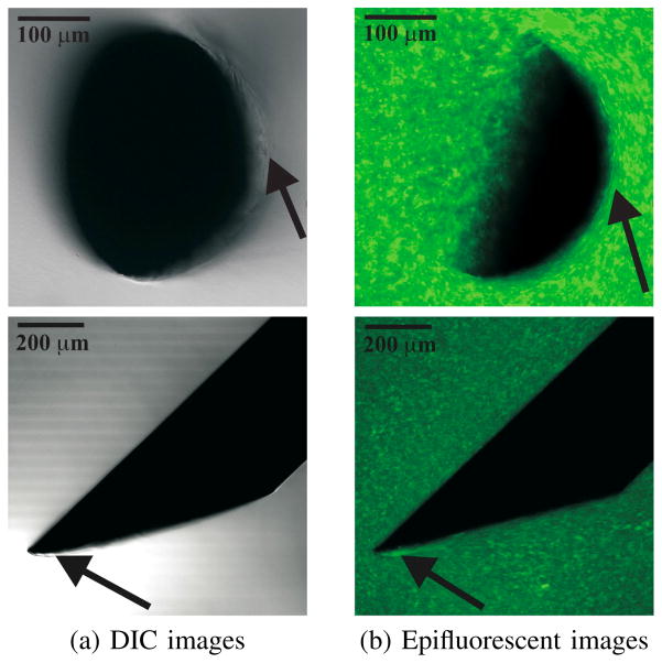
Fig. 3.
Example DIC and epifluorescent images taken using a confocal microscope. The top row of images are in the axial configuration, while the bottow row of images pertain to the perpendicular configuration. Needle geometric properties: Ø 0.40 mm and α = 33.9°. Arrows in the DIC images indicate that rupture of the gel is observed close to the bevel edge of the needle, while arrows in the epifluorescent images indicate that the gel is compressed near the needle shaft (axial) and the bevel face of the needle tip (perpendicular).

