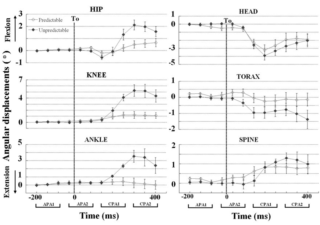
Figure 1.
Temporal evaluation (from −200 ms to + 400 ms in relation to T0) of the ankle, knee, hip, spine, thorax, and head displacements during predictable and unpredictable conditions. Each point represents the angular displacements in the sagittal plane (flexion (+) and extension (−) of these variables averaged over a 50 ms interval (−201 to −150 ms, −151 to −100, and so on) and its standard error. The 4 time epochs of 150 ms used for the analysis are represented by the brackets on the bottom (APA1, APA2, CPA1, and CPA2). The dotted vertical line shows the moment of body perturbation (T0).

