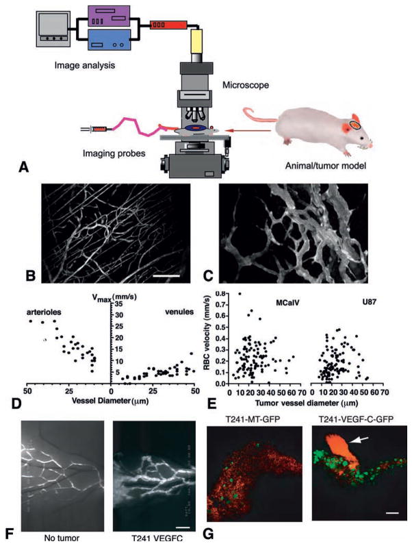
Fig. 1.
Imaging of tumor blood and lymphatic vessels. (A) Schematic intravital microscopy set up. An appropriate animal model, imaging probe(s), microscope, and image acquisition and analysis system are a prerequisite of intravital microscopy. (B–C) Multiphoton laser scanning microscopy image of normal blood vessels (B) and tumor vessels in LS174T human colon cancer xenografts (C) in mouse dorsal skin chambers. Blood vessels are contrast enhanced by FITC-dextran. Bar=100 μm. (D–E) Blood flow determined by intravital microscopy in normal pial vessels (maximum bead velocities, D) and tumor vessels in MCaIV murine breast cancer and U87 human glioma (RBC velocities, E) in mouse cranial windows. (F) FITC-dextran microlymphangiography of normal and peritumor lymphatics in mouse ears. Bar=850 μm. Lymphatic vessels are hyperplastic in the peritumor region and ear base (further downstream) of the T241 fibrosarcomas overexpressing VEGF-C. (G) GFP-positive tumor cells (green) are observed entering the cervical lymph node from afferent lymphatic (red, arrow) by MPLSM. Bar=100 μm. VEGF-C overexpression significantly increases arrival of tumor cells in the lymph node. Macroscopic lymph node metastasis increases with the increase of tumor cell arrival in the lymph node (58). B–C, courtesy of Dr. Edward Brown; D–E, adapted from (7); F–G, adapted from (58).

