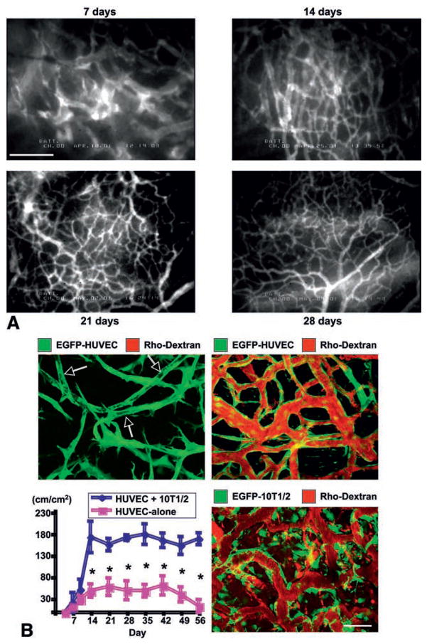
Fig. 3.
Imaging of physiological angiogenesis. (A) Angiogenesis and vessel remodeling during adipogenesis in the SCID mouse dorsal skinfold chamber after 3T3-F442A preadipocytes implantation. Blood vessels are contrast enhanced by the intravenous injection of FITC-dextran (2000 kDa). Bar=100 μm. (B) Morphological and functional analyses of engineered blood vessels. Vascular endothelial cells (HUVECs) and perivascular cell precursor cells (10T1/2 cells) or HUVECs alone were seeded in the collagen-fibronectin 3-D constructs and implanted in the cranial window in SCID mice. Top, 2-D projection of 3-D intravital multi-photon laser-scanning microscopy images of tissue-engineered blood vessels (EGFP expressing HUVEC, green; functional blood vessels contrast enhanced with rhodextran, red). Top left, 4 days after implantation of HUVEC+10T1/2 construct. Large vacuoles in the tubes resemble the lumens of capillaries (arrows) but they are not perfused (no red). Top right, 4 months after implantation of HUVEC+10T1/2 construct. Engineered vessels are stable and functional. Bottom left, Temporal changes in functional density of tissue-engineered blood vessels (total length of perfused vessel structure per unit area). N=4, mean ± SEM; *P<0.05. Bottom right, MPLSM image of engineered vessels, 4 weeks after implantation of HUVEC+EGFP-expressing 10T1/2 construct. Scale bar=50 μm. A, adapted from (101); B, adapted from (113).

