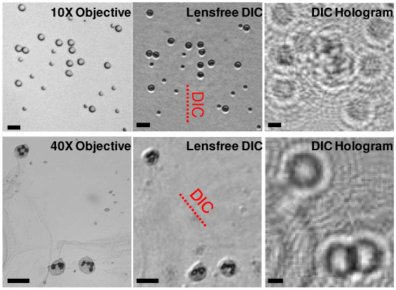
Fig. 2.

Reconstructed lensless DIC images of micro-objects. Top Row: 5 & 10 μm sized melamine (n = 1.68) beads in a medium (n = 1.524, Norland Optical Adhesive 65). The sample was illuminated at 550 nm (~18 nm FWHM bandwidth). A 50 μm aperture (at z1 = 10 cm) and 0.18 mm-thick quartz plate (δ ~1 μm) were used. Bottom Row: White blood cells in a blood smear sample are imaged. The sample was illuminated at 670 nm (~18 nm FWHM bandwidth) through a 50 μm aperture (z1 = 10 cm) and 0.3 mm-thick quartz plate (δ ~2 μm) was used for the DIC image. The shear directions are indicated in the figures with dashed lines. Conventional bright-field microscope images of the same FOV are presented for comparison. The scale bars are 20 μm.
