
Fig. 4.
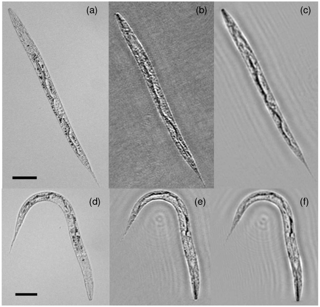
Caenorhabditis elegans imaging: (a) Microscope image (with a 10X objective lens – NA ~0.25); (b) Reconstructed lensfree DIC image; (c) Reconstructed regular lensfree holographic image; (d-f) same as (a-c), except for another C. elegans sample. Imaging conditions are the same as in Fig. 2. The scale bars are 50 μm.