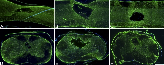
Fig. 3.
Immunohistochemical analysis with anti-glial fibrillary acidic protein (GFAP) (×4). At four weeks, the animals that had received a decompressive durotomy alone (B and E) displayed the most extensive astrocyte proliferation, indicating greater scarring relative to that in the contusion-only group (A and D) and that in the dural allograft group (C and F). Scale bar = 500 μm.

