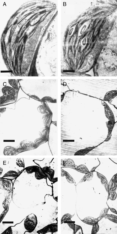
Fig. 3.
Transmission electron micrographs of chloroplast structure (A, B) and mesophyll cells (C–F) of irm1 and wild type: (A and B) chloroplast structure of young leaves of wild type (A) and irm1 (B); (C and D) an overview of a mesophyll cell of young leaves of wild type (C) and irm1 (D); (E, F) transmission electron micrographs of a mesophyll cell of wild type (E) and irm1 (F) grown on soil watered with Fe solution. Scale bar = 1 µm.

