Fig. 7.
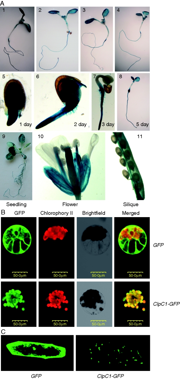
Expression pattern analysis of ClpC1 and subcellular localization of the ClpC1 protein. (A) Histochemical analysis of ClpC1 expression pattern using ClpC1 promoter-GUS transgenic lines: (1–4) GUS staining of the seedlings treated with Fe (2), Zn (3) or Mn (4) deficiency for 1 week; (5–8) GUS staining in the seedlings at 1, 2, 3 and 5 d after germination; (9–11) GUS staining of seedlings at the five-leaf stage and flowers and siliques from plants grown on soil. (B) Subcellular localization of ClpC1 by transient expression of ClpC1–GFP in cowpea protoplasts and in onion epidermis cells. The gene GFP (control) and ClpC1–GFP (a fusion gene of ClpC1 and GFP) under control of the 35S promoter were separately introduced into cowpea protoplasts using PEG transformation and expressed. (C) GFP (control) and ClpC1–GFP were transiently expressed in onion epidermis cells introduced by bombardment. The GFP signal is localized in chloroplasts of cowpea protoplasts (B) and in plastids of onion epidermis cells (C) when transformed with 35S::ClpC1–GFP, whereas the GFP signal was spread through the whole cytoplasm in the cells that were transformed with 35S::GFP. Scale bar = 50 µm.

