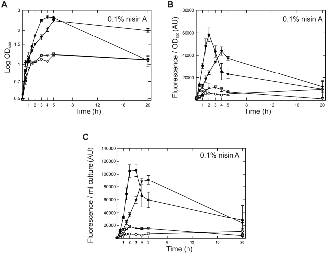
Figure 2. Time-resolved protein expression in GM17- and GCDM-grown cells.
(A) Growth of L. lactis NZ9000 in GM17 (closed symbols) and GCDM (open symbols), following the addition of 0.1% of nisin A-containing NZ9700 medium supernatant to a culture at OD600≈0.5. The cells express either OpuAC-GFP (circles) or BcaP-GFP (inverted triangles). Growth of the cells was monitored by measuring the optical density at 600 nm. (B and C) Time dependence of OpuAC-GFP and BcaP-GFP expression (symbols the same as in panel A). Expression levels were quantitated on the basis of GFP fluorescence and normalized on the basis of cell density (panel B) or culture volume (panel C).

