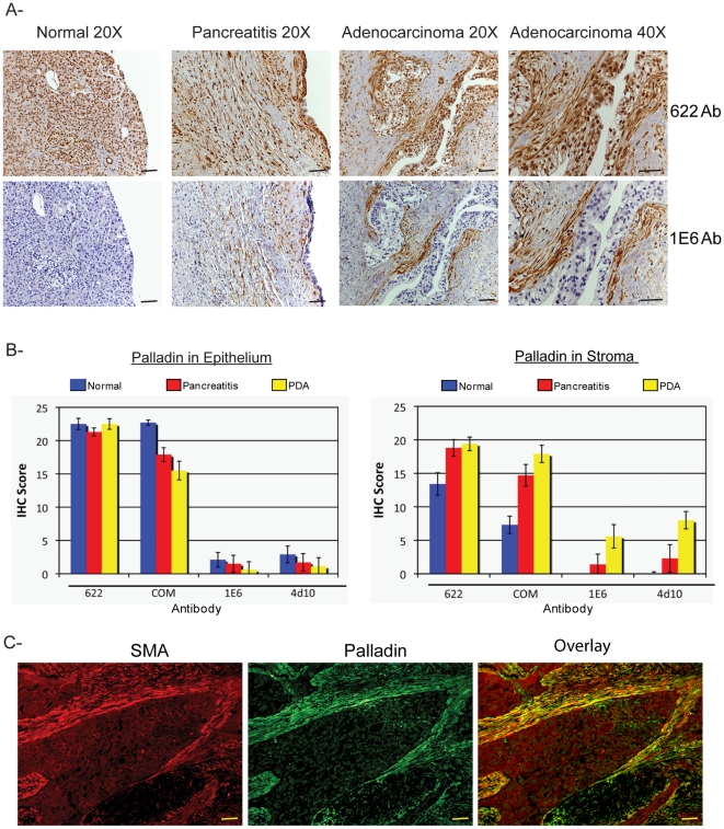
Figure 3. Palladin staining of paraffin-embedded patient tissues.
A. IHC staining was performed using standard antigen-retrieval protocols, and counter-stained with hematoxylin. Tissue sections were stained for palladin using two palladin antibodies: polyclonal 622 and monoclonal 1E6. Palladin stain is detected with brown reaction product. In tumor sections, palladin is detected at dramatically elevated levels in the stromal fibroblasts. Note also the expanded stroma around the neoplastic cells, which is characteristic of the desmoplastic reaction. Scale bars, 200 µM. B. Quantification of immunohistochemistry results. Ten sections each of normal pancreatitis and adenocarcinoma specimens were stained with four different antibodies (622, COM, 1E6 and 4D10) and scored by two pathologists, as described in the text. Results for both ductal epithelium (left) and stroma (right) stained with various palladin antibodies are shown for normal pancreas (n = 9, blue), pancreatitis (n = 7, red) and pancreatic adenocarcinoma (n = 10, yellow). The results confirmed that palladin levels are increased in the stroma, and not the epithelial tumor cells, of the adenocarcinomas. Although palladin levels are also increased in cases of chronic pancreatitis, they do not reach the same levels as in the tumors. Compared to the polyclonal 622 and COM, the monoclonal antibodies 1E6 and 4D10 are effective at distinguishing between pancreatitis and cancer. C. Double-label immunostaining for palladin (1E6 Ab) and α-SMA in sections of pancreatic tumors confirms that palladin is strongly detected in a population of activated TAFs that surround the neoplastic cells. Scale bars, 200 µM.

