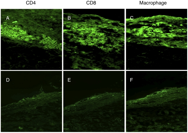
Figure 5. Cellular infiltration in rejecting and tolerant grafts.
Cryostat sections were stained by anti-mouse CD4 (A–D), CD8 (B–D) and macrophage antibodies (C–E) in Group 1 (A–B–C) and Group 4 at 200 days post transplantation. In Group 1 of rejecting mice, immunohistology for cellular immune responses to concordant islet xenografts at time of rejection has detected mixed cellular infiltrates with presence of CD4+ (A), CD8+ (B), and macrophages (C). In Group 4 of tolerant mice, immunohistology for cellular immune responses to concordant islet xenografts of tolerant mice at 200 days post-transplantation detected only minimal cellular infiltrate with absence of CD4+ (D), CD8+ (E), and macrophages (F). (Magnification in A–C (200x), Magnification in D–F(100x)).

