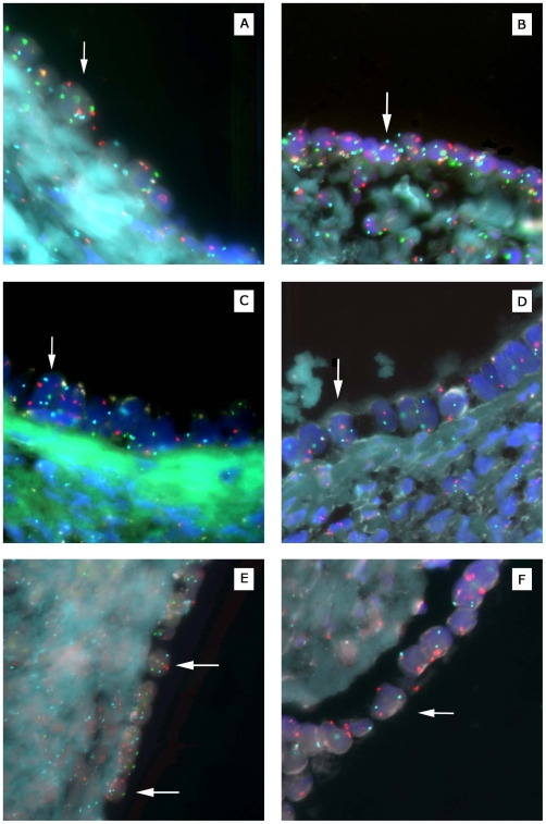
Figure 4. Fluorescence in situ hybridization analysis of ploidy in normal ovarian cystic and surface epithelia.
All examples shown are from distinct ovarian specimens. (A) Cystic epithelial cell (arrow) from sample #112 (see Table 2) with three copies of chromosomes 3 (red) and 11 (green). (B) Cystic epithelial cell from sample #84 with three copies of chromosomes 6 (blue) and 11 (green). (C) Cystic epithelial cell from sample #135 with three copies of chromosome 6 (blue). (D) Cystic epithelial cell from sample #59 with three copies of chromosome 8 (blue). (E) Surface epithelial cells from sample # 13 with two copies of chromosomes 3 (red), 6 (blue), and 11 (green). (F) Surface epithelial cell from sample #102 with two copies of chromosomes 3 (red) and 8 (blue).

