
Figure 3. Inflammatory cells in thoracic and abdominal aortic aneurysms.
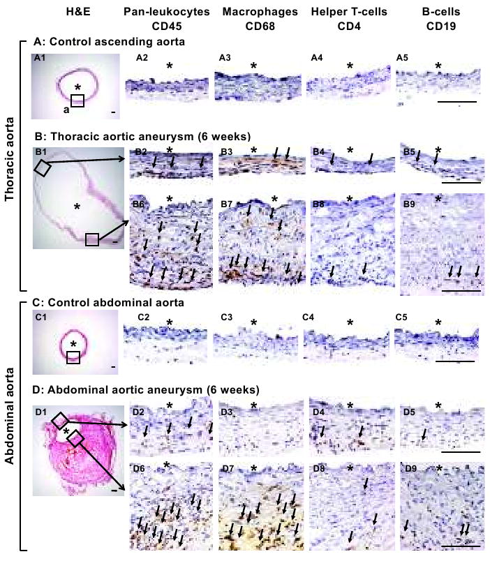
Stainings for pan-leukocytes (CD45), macrophages (CD68), helper T-lymphocytes (CD4) and B-lymphocytes (CD19). In thoracic aneurysms, leukocytes were observed in the adventitia and media (B2, B6). In contrast, leukocytes were highly concentrated in the thick wall near the intramural thrombus in abdominal aortic aneurysms (D6). In both thoracic and abdominal aneurysms, majority of leukocytes appeared to be macrophages (B2-5, B6-9, D2-5, D6-9). *: lumen. Scale bar: 0.1mm. Arrows point to positive cells.