
Figure 1. A: PPARα, β/δ, and LXR Activators Reverse Murine Ox-AD.
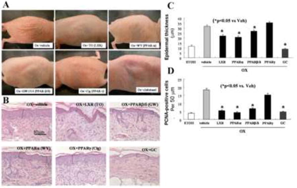
Gross appearance after applications of ligands for LXR(10mM T0901317), PPARα(10mM WY14643), β/δ(4mM GW1514), and γ (10mM ciglitazone), and glucocorticoid (GC; 0.05% clobetasol). As a vehicle control (Ox+vehicle), propylene glycol and ethanol (7:3) alone was applied. B: Histological appearance after treatment with ligands for LXR, PPARα, β/δ and γ, and the glucocorticoid (GC), clobetasol. (H&E staining) C: Quantitative changes in epidermal hyperplasia, in LXR, PPARα, β/δ, and γ ligands, and the glucocorticoid (GC), clobetasol - treated mice were assessed in coded, randomized micrographs (see Methods). D: PCNA-positive cells counts (per 50 μm) were quantitated as described in Methods. ETOH: Normal skin in which ethanol was applied instead of Ox (n=30 measurements each from 3 separate samples for epidermal hyperplasia assessment; and 22-27 measurements each for PCNA assessment).