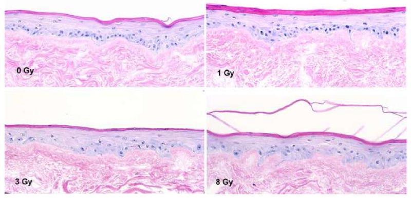
Figure 3.

The histological change of EVPOMEs after ionizing radiation. At 0 Gy, the EVPOME appeared healthy with well aligned cells along the basement membrane. At 8 Gy, oral mucosa keratinocytes were aberrant in alignment and faint nuclear staining of the basal cells was seen. The basal cell had pyknoic nuclei. (HE stain, ×200)
