Figure 4.

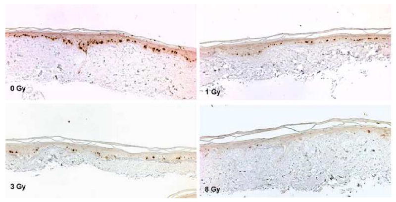
The result of Ki-67 immunohistochemistry. At 0 Gy, Ki-67 positive cells are mainly seen within the basal layer of cells. At 8 Gy, the number of positive cells dramatically decreased and they were scarce (×200).

The result of Ki-67 immunohistochemistry. At 0 Gy, Ki-67 positive cells are mainly seen within the basal layer of cells. At 8 Gy, the number of positive cells dramatically decreased and they were scarce (×200).