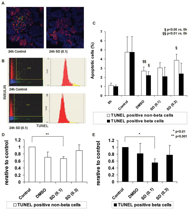
Fig. 2. β cell apoptosis in cultured human islets is prevented by supplementing the medium with 0.1 μM SD-282 as determined by Laser Scanning Cytometry.
Islet sections were labeled by TUNEL to identify apoptotic cells, immunostained for insulin to identify β cells, and quantified using laser scanning cytometry. A: Representative merged images of islets cultured for 24 hours (left – medium alone (control), right – medium supplemented with 0.1 μM SD-282). Green – TUNEL-positive, red – insulin-positive, yellow – TUNEL-insulin double-positive, and blue – DAPI DNA-staining; B: Scattergrams and histograms obtained by scanning the adjacent slides; C: Percentages of apoptotic cells apoptotic β cell percentages were calculated by dividing the TUNEL-insulin double-positive cell number by the total insulin-positive cell number in each section. TUNEL-positive/insulin-negative cells represent apoptotic non-β cells. Percentages of non-β cells were calculated by dividing the TUNEL-positive/insulin-negative cell number by the total number of non-β cells. After 24 hours culture, the mean apoptotic non-β cells (%) significantly increased in the DMSO (p<0.01) and 0.3 μM SD-282 (p<0.05) groups as compared to pre (0h). The mean apoptotic β cells (%) significantly increased in the DMSO (p<0.05) group as compared to pre (0h). To determine SD-282 effects, the relative ratio was obtained by dividing the post-culture apoptotic cell percentage by that of the corresponding medium alone control group; D: Relative ratios of apoptotic non-β cells – Culture medium supplemented with 0.1 μM SD-282 significantly prevented non-β cell apoptosis as compared to control (p<0.001); E: Relative ratios of apoptotic β cells – β cell apoptosis was significantly decreased with 0.1 μM SD-282 in the medium (p<0.01). This SD-282 effect was not observed at a concentration of 0.3 μM (n=5).

