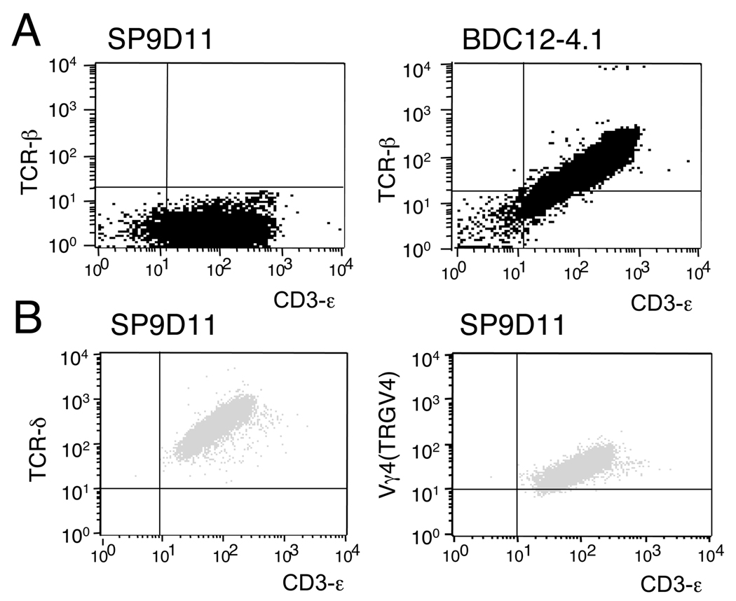
Figure 1. The SP9D11 hybridoma: absence of TCR-β expression.
A: SP9D11 cells and an ins2 B:9–23 reactive TCRαβ+ hybridoma (BDC12-4.1) were stained with mAbs specific for TCR-β and CD3ε, and analyzed by flow cytometry. B: SP9D11 cells were stained with mAbs specific for TCR-δ or TCR-Vγ4 (TRGV4) and CD3ε, and analyzed by flow cytometry. Data shown are representative of multiple (>3) independent staining experiments.

