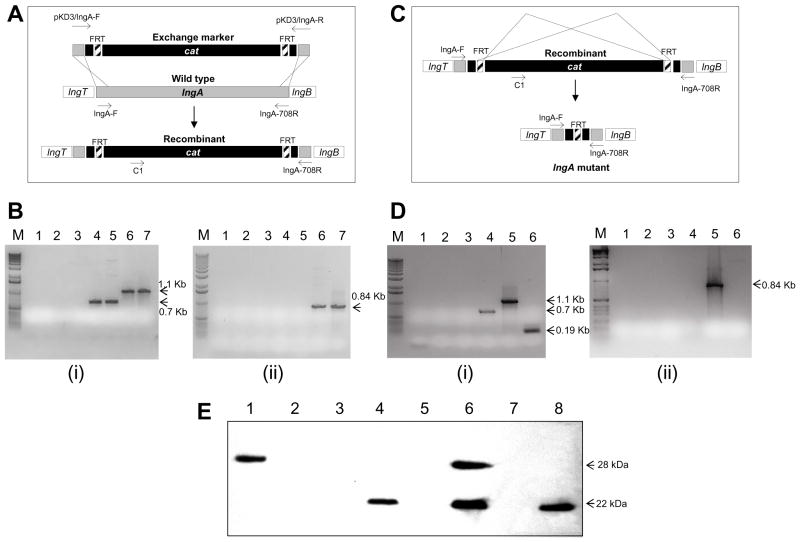
Figure 2. Deletion mutation of the lngA gene.
Panel A. Diagram of the cat cassette insertion into the lngA gene by homologous recombination. Diagram of the wild-type lngA gene and a recombinant lngA:cat DNA fragment that by homologous recombination event result in the lngA:cat insertion mutant. Primers pKD3-lngA-F/pKD3-lngA-R to construct lngA::FRT-cat exchange marker and primers lngA-F/lngA-708R to test for lngA sequences are shown. Panel B. Detection of lngA:cat DNA recombination by PCR amplification. i) DNA PCR amplification for detection of lngA DNA using primer set lngA-F/lngA708-R. Lane 1: E. coli DH5α; Lane 2: DH5α pKD46; Lane 3: DH5α pKD3; Lane 4: E9034A; Lane 5: E9034A(pKD46); Lane 6: DH5α pAClngA:FRT-cat; Lane 7: E9034AlngA:FRT-:cat. Arrows indicate DNA fragments of 1.1 and 0.7 kb. ii) DNA PCR amplification for detection of cat insertion using primer set C1-lngAR PCR. Lanes 1 to 7: same as panel B-I above. Arrow indicates a 0.84-Kb DNA fragment. Panel C. Diagram the isogenic lngA deletion mutation construction, showing the Flp-mediated cat cassette excision by homologous recombination at the FRT sites. The resulting lngA deletion mutant contains an FRT site in the middle of lngA. Primer c1/lngA-708R for recognition of cat sequences and primers lngA-F/lngA708R for detection of lngA sequences are shown. Panel D. Detection of lngA deletion mutation by DNA PCR amplification. i) PCR amplification for detection of lngA DNA using primer set lngA-F-lngA708R. Lane 1: DH5α; Lane 2: DH5α pCP20; Lane 3: E9034A; Lane 4: E9034AlngA::FRT-cat; Lane 5: E9034AlngA::FRT-cat(pCP20); Lane 6: E9034AΔlngA. Arrows indicate 1.1, 0.7, and 0.19-Kb DNA fragments. ii) PCR amplification for detection of cat DNA using primers C1-lngA708-R. Lanes 1 to 6: same as panel D-i above. Arrow indicates a 0.84-Kb DNA fragment. M represents molecular weight markers (Bioline, Tauton, MA). Panel E. Expression of LngA. The expression of LngA was determined by immunoblot in whole cells lysates using a anti-Longus monoclonal antibody. Lane 1, E coli DH5α(pAClngA); Lane2, E coli DH5α; Lane 3, E coli HB101; Lane 4, E9034A; Lane 5, E9034AΔlngA; Lane 6, E9034AΔlngA(pAC-lngA); Lane 7, E9034AΔlngA::FRT-cat; Lane 8, E9034AΔlngA::FRT-cat (pAC-lngA). Arrows indicate the 28 kDa LngA prepilin and the 22 kDa mature LngA pilin.

