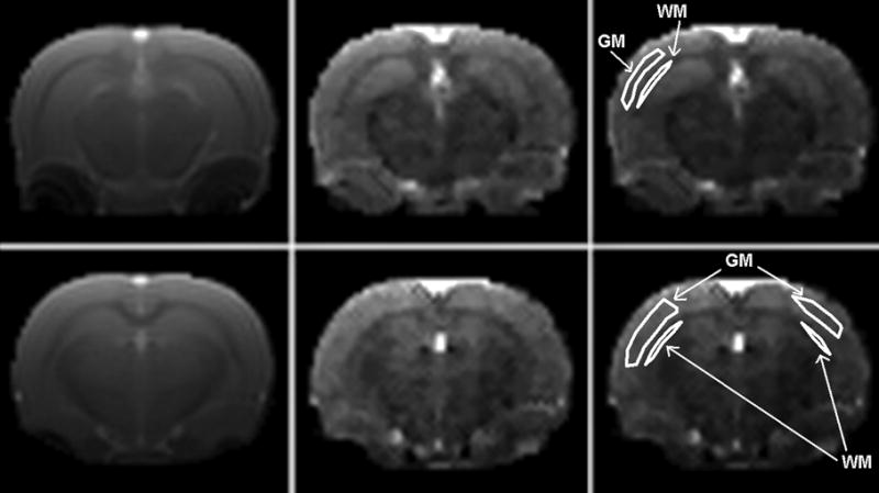
FIGURE 3.

Representative brain ASL images acquired using the FAIR-TrueFISP with QUIPSS sequence with non-selective inversion (left column), and corresponding CBF maps (central column). Right column is the replicated CBF map showing representative ROI drawings for the gray (GM) and white matter (WM) definition. Imaging parameters were: matrix size = 128×128, FOV = 35 × 35 mm, slice thickness = 2.0 mm, TR = 2.68 ms, TE = 1.34 ms, TI = 1400 ms, ΔTI (QUIPSS) = 700 ms, slab thickness (inversion) = 12 mm, and repetition time for inversion = 5000 ms.
