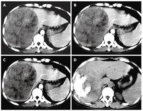
Figure 2.

Transverse CT scans showing a massive pattern of HCC in the right lobe of a 46-year-old man with a tumor volume of 1936.70 cm3, a TTLVR of 67.52%, and a survival time of 6 mo. A and B: Arterial phase contrast- enhanced CT scan showing a high density tumor in the right lobe of liver before the first TACE procedure, while one-dimensional measurement showing the largest tumor diameter (17.7 cm) and two-dimensional measurement showing the tumor product of the greatest axial dimension (237.18 cm2); C: Contrast-enhanced CT scan showing hepatic lesion volumetric measurements before TACE; D: Nonenhanced CT follow-up scan showing lipiodol retention of grade III and a reduced tumor size 60 d after the first TACE procedure.
