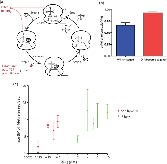
Figure 4.
In vitro termination assays reveal that Ribo-X has a decreased affinity for RF1. (a) Schematic of termination assays. A complex of the O-ribosome with an initiator tRNA aminoacylated with 35S formyl-methionine (red circle) is assembled (Step 1), in subsequent rounds this requires dissociation of deacetylated tRNA. RF1 is added (Step 2) directing hydrolysis of 35S formyl-methionine from the tRNA (Step 3). RF1 dissociates (Step 4). The filter-binding assay measures the ribosome associated 35S, while the TCA precipitation measures the 35S released (b) RF1 termination activity measured in vitro with untagged wild-type and purified tagged O-ribosome, using filter-binding assays. (c) O-ribosome and Ribo-X termination activity with RF1. Measurement of initial rate of tRNA-fMet hydrolysis over a range of RF1 concentrations is shown for O-ribosome and Ribo-X. RF1 concentration was increased till there was a measurable release in fMet and this release plotted. tRNA-fMet hydrolysis (37°C, 10 s) is measured by TCA precipitation.

