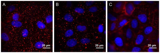
Figure 5.
Epi-fluorescence microscopy (Olympus IX71, 60X, 1.20 N.A. objective) was used to image cells following a 4 min incubation at 37°C. DAPI (blue) was used as a nuclear stain. (a) 4 nM PA-QDs (red) in the presence of 4 μM pyrenebutyrate (positive control). (b) 4 nM PA-QDs (red) in the absence of pyrenebutyrate (negative control). (c) 4 nM QDs (red), in the absence of PA, in the presence of 4 μM pyrenebutyrate (negative control).

