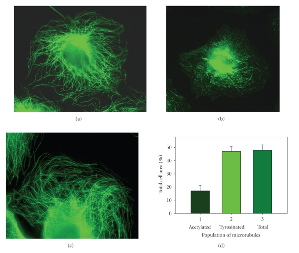
Figure 1.
Dynamic microtubules are directed towards and their ends reach the cell periphery. HPAEC were fixed and processed for immunofluorescence microscopy. (a) Antibodies against β-tubulin were used to detect total microtubule population of the cell; (b) antibodies against acetylated tubulin were used to detect stable microtubules; (c) antibodies against tyrosinated tubulin were used to detect dynamic microtubules. Scale bar, 20 μm; (d) relative area occupied by microtubules (% of the total cell area): 1—acetylated microtubules; 2—tyrosinated microtubules; 3—total population of cell microtubules.

