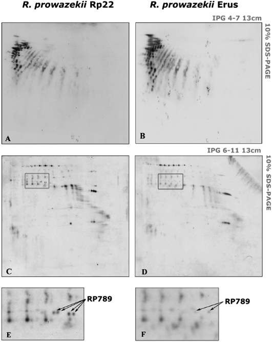
Figure 6.
Lysine methylation in Rp22 (A,C,E) and Erus (B,D,F) strains. Two-dimensional Western blots were performed, and lysine methylation was visualized in pH range 4.0–7.0 (A,B) and pH range 6.0–11.0 (C,D). The zone that differed among both bacterial strains is boxed in C and D. Arrows in E and F indicate methylated spots of Rp22 and Erus strains that were identified as putative methyltransferase (RP789).

