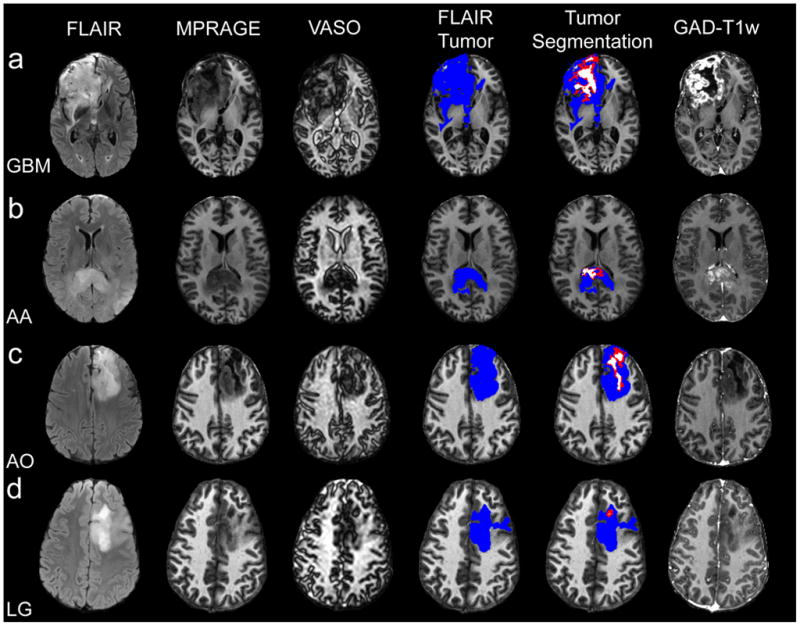
Figure 4.

One representative slice for a GBM (a), AA (b), AO (c) and low-grade (LG) (c). Long-T1 zones exist, to varying spatial extents, in the high-grade tumors, with negligible detectable long-T1 zone in the low-grade tumor. Notice the dark boundary surrounding the VASO hyperintense (long-T1) zone corresponding to a non-enhancing central tumor area, which is putatively assigned to necrosis (see discussion).
