
Figure 2.
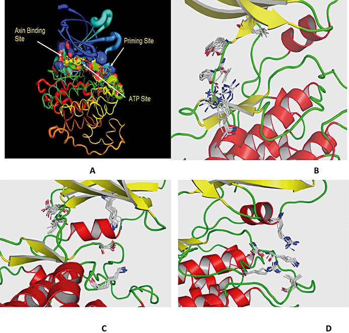
Different binding pockets of GSK3β. (A) Overlay of X-ray structures at Cα depicting different binding sites for GSK3β; (B) ATP site; (C) axin binding site; (D) priming site. The residues of each site are overlaid to understand the mobility of each residue. The helices are denoted in red, β-sheets in yellow and loops in green colour. The residues are shown in elemental colours. GSK3β, glycogen synthase kinase-3 beta.