Figure 6.

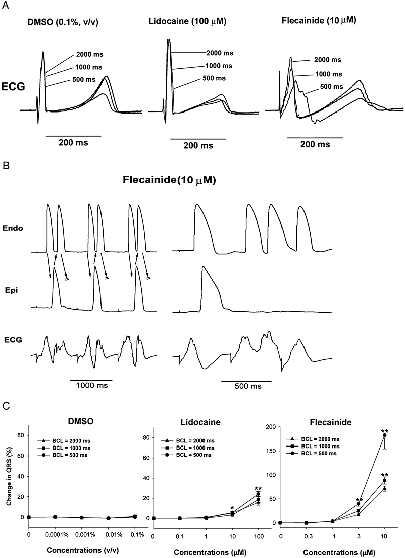
Effects of lidocaine (100 µM) and flecainide (10 µM) in the isolated rabbit left ventricular arterially perfused wedge preparation. (A) The effects of solvent (DMSO), lidocaine (100 µM) and flecainide (10 µM) on ECG at stimulation cycle lengths of 2000, 1000 and 500 ms. Note that lidocaine weakly increased QRS duration in a non-rate-dependent manner, while flecainide strongly increased QRS duration in a rate-dependent manner. (B) Flecainide-induced ectopic beats and VT. Note that the slowing of conduction between the endocardial (Endo) and epicardial (Epi) layers caused re-entrant beats, that is, extra-systoles (left panel) and three beats of VT (right panel). Stimulation at basic cycle length = 500 ms. (C) The effects of solvent (DMSO), lidocaine and flecainide on QRS duration. Note that lidocaine slightly, but significantly, increased QRS duration (slowing of conduction), and flecainide markedly increased QRS duration in a concentration-dependent and a rate-dependent manner (n= 7 per group). **P < 0.01, significantly different from effects of solvent.
